MICSI-RMT is the first introduction of the popular MP-PCA algorithm into clinical practice. The method utilizes a fast-search algorithm to detect and discard noise-only components that are defined using the Marchenko-Pastur distribution.
The method exploded in popularity for two reasons, (1) its accuracy in removing noise while preserving the signal and (2) its denoising power.
MP-PCA acts as an intelligent averaging tool that combines the many images from the MRI exam, preserving their unique image properties and discarding noise. The degree of image enhancement scales with the number of images in the dataset.
MP-PCA does not hallucinate.[1, 2, 6]
The principal component basis enables precise
separation between signal and noise, thereby preserving anatomy during denoising.
The performance of the algorithm does not depend on externally trained datasets. This approach is self-supervised, meaning that only the images from exam would be used for training and denoising.
All other denoising approaches—filters, wavelets, or even the latest AI neural networks—work by executing weighted combinations of various regions within the image. This process essentially disperses the signal across different parts of the image, leading to loss of signal in addition to noise.
MP-PCA uses the different images over the 4D stack to learn the noise level and remove the components related to noise. If the algorithm fails to detect the noise components, then no denoising occurs, thereby ensuring that this algorithm removes noise without eliminating vital anatomy.
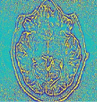
The comparison shows the original, MP-PCA, 3-D Gaussian Filter, Anatomical Non-Local Means (ANLM),
and a convolutional neural network trained for denoising in 2D. The residuals are plotted for each
method to highlight which regions were altered with each denoising method.
MP-PCA is
the only method that does not remove anatomy in the brain.[1, 2, 6]
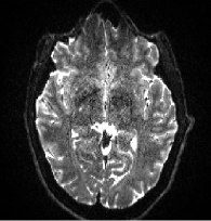
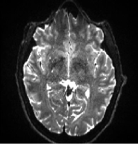
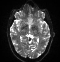
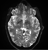



MP-PCA can be conceptualized as a smart averaging approach. It combines images with different contrasts, time-points, orientations, or motion states to eliminate noise, as if these images were averaged together. This eliminates the need for in-line averaging and encourages the acquisition of more measurements to increase the information content of the exam. This method fosters a new approach to imaging: acquire as many images with different contrasts as possible (regardless of their SNR), then use them in combination to achieve a significant boost in SNR.
MP-PCA adds a new term to the SNR equation for MRI measurements. This term enables a multiplicative boost in SNR-based on the number of measurements across the entire image series.

MP-PCA revolutionizes diffusion MRI by enabling high diffusion weightings, or b-values. In high b-value images, the signal throughout the brain is exponentially suppressed, leading to incredibly noisy original images. The suppression is greater for structures that have less restrictions to diffusion of water, such as signals from cerebrospinal fluid and extra-axonal spaces. With MP-PCA it is possible to produce clear, crisp images at high diffusion weightings, unlocking insights into the intricate properties of the brain’s intra-axonal regions.
Most Cited Paper on Diffusion MRI
1350+ citations from 20+ countries
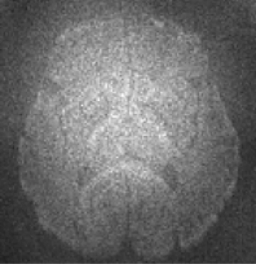
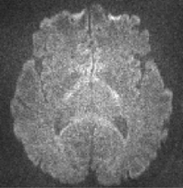
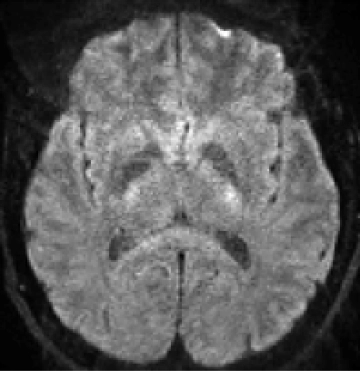
The same patient was imaged with different scanners.
MP-PCA enables high image quality at
all resolutions and bridges the gap in image quality between low-end and high-end systems.
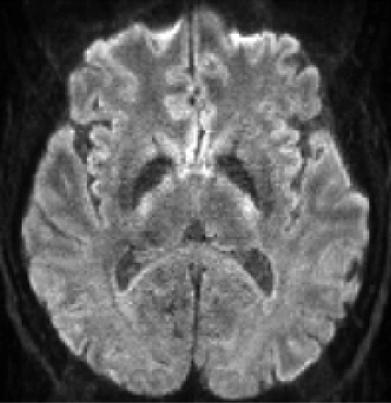
Using MP-PCA, patients reach the same sensitivity level (number of voxels with z > 3) in just 60% of the original imaging time for finger- tapping & verb-generation tasks.


By registering to download the brochure, you are consenting and agreeing to collection and use of your information by MICSI in accordance with its Privacy Policy.